Free 
3D model · NIH 3D
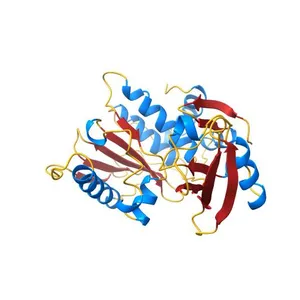
Crystal structure of CYLD USP domain (C596S) in complex with Met1-linked diubiquitin
Model details
- Platform
NIH 3D
- Price
- Free
- License
- CC0
- Public domain: print, modify and sell, no attribution required.
- Formats
- X3DGLBWRLSTL
- 8 Downloads
- 125 Views
Description
PDB Classifiation: HYDROLASE/PROTEIN BINDING
Similar models
Free 
The crystal structure of human Atg4B(C74S)- LC3(1-124) complex
Free 
Structural insights into the interaction of p97 N-terminal domain and SHP motif in Derlin-1 rhomboid pseudoprotease
Free 
Crystal structure of thrombin mutant S195T (free form)
Free 
Crystal Structure of Calcineurin in Complex with the Calcineurin-Inhibiting Domain of the African Swine Fever Virus Protein A238L
Free 
Crystal Structure of Multi-drug Resistant HIV-1 Protease PR-S17 in Complex with Darunavir
Free 
Crystal Structure of sv40 large T antigen origin binding domain with DNA
Free 
Crystal structure of the USP7/HAUSP catalytic domain
Free 
Crystal structure of the C276S mutant of MAP2K7
Free 
Crystal structures of aldehyde deformylating oxygenase from Oscillatoria sp. KNUA011
Free 
Structure of Florigen Activation Complex Consisting of Rice Florigen Hd3a, 14-3-3 Protein GF14 and Rice FD Homolog OsFD1
Free 
Structural Details of Ufd1 binding to p97
Names are used only to indicate the source of a result. Polyzon is not affiliated with the listed sites.