Free
3D model · NIH 3D
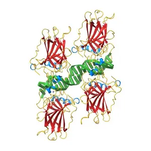
Crystal structure of the human p73 tetramerization domain
Model details
- Platform
NIH 3D
- Price
- Free
- License
- CC-BY
- Print, modify and use commercially — credit the author.
- Formats
- WRLGLBSTLX3D
- 8 Downloads
- 152 Views
Description
PDB Classifiation: TRANSCRIPTION
Similar models
Free
Crystal structure of human TBX5 in the DNA-bound and DNA-free form
Free
Human p53 core domain with hot spot mutation R249S and second-site suppressor mutations H168R and T123A
Free
Crystal structure of human TBX5 in the DNA-free form
Free
p53 cancer mutant Y220C
Free
Structure of the FOXM1 DNA binding
Free
Human p53 core domain with hot spot mutation R273C
Free
Crystal structure of human YAP and TEAD complex
Free
Crystal Structure of Rhodobacter sphaeroides SigE in complex with the anti-sigma ChrR
Free
humanRXR alpha & mouse LXR alpha complexed with Retenoic acid and GSK2186
Free
Crystal structure of the human PARP-1 DNA binding domain in complex with DNA
Free
Lysine 120-acetylated P53 DNA binding domain in a complex with DNA.
Names are used only to indicate the source of a result. Polyzon is not affiliated with the listed sites.
Comments