Free
3D model · NIH 3D
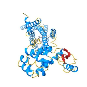
Crystal structure of human GLUT3 bound to D-glucose in the outward-occluded conformation at 1.5 angstrom
Model details
- Platform
NIH 3D
- Price
- Free
- License
- CC-BY
- Print, modify and use commercially — credit the author.
- Formats
- STLWRLGLBX3D
- 11 Downloads
- 304 Views
Description
PDB Classifiation: TRANSPORT PROTEIN
Similar models
Free
Crystal structure of the human glucose transporter GLUT1
Free
Crystal Structure of Human Galectin-3 CRD in Complex with Type 1 N-acetyllactosamine
Free
The Crystal Structure of Human Argonaute2 Bound to a Guide and Target RNA Containing Seed Pairing from 2-8
Free
Crystal structure of the 5-HT2B receptor solved using serial femtosecond crystallography in lipidic cubic phase.
Free
Crystal structure of the Bovine Fructose transporter GLUT5 in an open inward-facing conformation
Free
Crystal structure of GluN1/GluN2B NMDA receptor, structure 1
Free
Crystal structure of CC2-SUN of mouse SUN2
Free
Crystal structure of Human galectin-3 CRD in complex with lactose (pH 7.5, PEG6000)
Free
Crystal Structure of Myosin-1c tail in complex with Calmodulin
Free
The structure of the MFS (major facilitator superfamily) proton:xylose symporter XylE bound to D-glucose
Free
Crystal Structure at 3.4 A Resolution of Fully Glycosylated HIV-1 Clade G X1193.c1 SOSIP.664 Prefusion Env Trimer in Complex with Broadly Neutralizing Antibodies PGT122, 35O22 and VRC01
Names are used only to indicate the source of a result. Polyzon is not affiliated with the listed sites.