3D model · NIH 3D
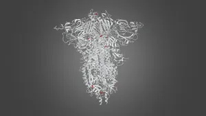
SARS-CoV-2 spike protein 6VSB with one Receptor Binding Domain (RBD) up in the active conformation
Model details
- Platform
NIH 3D
- Price
- Free
- License
- CC-BY
- Print, modify and use commercially — credit the author.
- Formats
- X3DGLBSTLWRL
- 23 Downloads
- 258 Views
Description
SARS-CoV-2 spike protein from 6VSB. The protomer that has its Receptor Binding Domain (RBD) flipped into the up (active) conformation has its domains colored as described in Figure 1 of: Wrapp, D. , Wang, N. , Corbett, K.S. , Goldsmith, J.A. , Hsieh, C.L. , Abiona, O. , Graham, B.S. , McLellan, J.S. (2020) Science 367 : 1260-1263
Blue: N-Terminal Domain (NTD)
Green: Receptor Binding Domain (RBD)
Tan: Remainder of S1
Cyan: Fusion Peptide
Yellow: Heptad Repeat 1
Orange: Central Helix
Purple: Connector Domain
Red: Remainder of S2
See also 3dpx-012877 for a related structure from SARS-CoV with all RBD down in the inactive conformation.
Similar models
Free
Free
SARS-CoV-2 Spike S Protein and ACE2 (PathogenAR)
Free
SARS-CoV-2 Open Spike (S-Protein)
Free
SARS-CoV-2 Spike Protein Molecular Structure
Free
Structure of sybody MR17-K99Y in complex with the SARS-CoV-2 S Receptor-binding domain (RBD)
Free
Structure of sybody MR17 in complex with the SARS-CoV-2 S receptor-binding domain (RBD)
Free
SARS-CoV-2 Open Spike (S-Protein) with Ab fragment (PathogenAR)
Free
SARS-CoV-2 Spike protein, Smithsonian Futures Exhibit
Free
Crystal structure of receptor binding domain of SARS-CoV-2 Spike glycoprotein in complex with EY6A Fab
Free
SARS-CoV-2 Open S Protein (Spike Protein) in Membrane (PathogenAR)
Free
Structure of sybody SR4 in complex with the SARS-CoV-2 S Receptor Binding domain (RBD)
Free
SARS CoV-2 Virion (NIAID)
Names are used only to indicate the source of a result. Polyzon is not affiliated with the listed sites.
Comments