Free 
3D model · NIH 3D
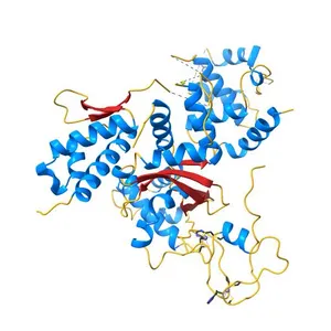
Structure and Function of a Ubiquitin Binding Site within the Catalytic Domain of a HECT Ubiquitin Ligase
Model details
- Platform
NIH 3D
- Price
- Free
- License
- CC-BY
- Print, modify and use commercially — credit the author.
- Formats
- GLBWRLX3DSTL
- 5 Downloads
- 148 Views
Description
PDB Classifiation: LIGASE
Similar models
Free 
Biotin protein ligase R40G mutant from Aquifex aeolicus in complex with biotin
Free 
Crystal structure of the HPV16 E6/E6AP/p53 ternary complex at 2.25 A resolution
Free 
Structure of active beta-arrestin1 bound to a G protein-coupled receptor phosphopeptide
Free 
Crystal structure of the THAP domain from D. melanogaster P-element transposase in complex with its natural DNA binding site
Free 
Crystal structure of the USP7/HAUSP catalytic domain
Free 
Crystal structure of a designed Mn binding peptide
Free 
HA17-HA33-Lac
Free 
Crystal structure of the Vif-CBFbeta-CUL5-ElOB-ElOC pentameric complex
Free 
X-ray crystal structure of a heavy metal efflux pump, crystal form I
Free 
Crystal structure of the auto-inhibited Nedd4 family E3 ligase Itch
Free 
Crystal structure of human prolyl-tRNA synthetase (halofuginone bound form)
Names are used only to indicate the source of a result. Polyzon is not affiliated with the listed sites.