
Free 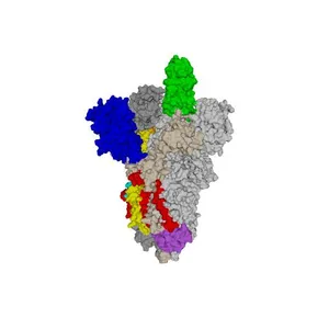
SARS-CoV-2 spike protein 6VSB with one Receptor Binding Domain (RBD) up in the active conformation
NIH 3D
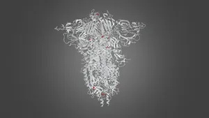
SARS-CoV-2 Spike glycoprotein model displayed in the Smithsonian Institution Futures exhibit. The Spike glycoprotein projects from the outer surface of the SARS-CoV-2 virus and is the basis for COVID-19 vaccines. “Glycoprotein” means that it is mostly made of protein with attached sugars (but the sugars are not included on this model). This structure represents a complete Spike protein, and is a mixture of experimental and computer modeled data.
This structure is made up of 3 identical protein chains, two of them are shown here as red and blue molecular surfaces and the third is shown as a ribbon that indicates the path that the protein backbone takes under the surface with rainbow coloring varying from the start to the end of the protein chain.
The bottom is called the stem, and it inserts into the membrane on the outer surface of the virus particle. The cyan color on the stem corresponds to the location of the outer part of the viral membrane, with the inner part of the membrane just above the wider knob at the bottom. The wider top is called the head, and it attaches the virus to receptors on the outer surface of human cells to start the infection process. Note that the top of the red surface has a section, the Receptor Binding Domain (RBD) that has flipped “up”; this position is required to bind to the receptors. The other two chains have the RBD in the “down” position which protects the virus from the immune system.
The magenta color near the upper center of each of the 3 parts shows the “2P” mutations which were engineered to stabilize the Spike; this stabilization is what makes the vaccines possible.
This model was prepared at the direction of Dr. Barney Graham, the former Deputy Director of the Vaccine Research Center of the National Institute of Allergy and Infectious Diseases.

Names are used only to indicate the source of a result. Polyzon is not affiliated with the listed sites.