Free 
3D model · NIH 3D
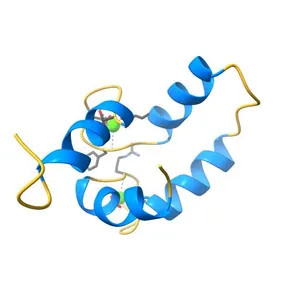
NMR structure of the dilated cardiomyopathy mutation G159D in troponin C bound to the anchoring region of troponin I
Model details
- Platform
NIH 3D
- Price
- Free
- License
- CC-BY
- Print, modify and use commercially — credit the author.
- Formats
- GLBX3DWRLSTL
- 78 Downloads
- 46 Views
Description
PDB Classification: METAL BINDING PROTEIN
Similar models
Free 
Solution NMR structure of the C-terminal EF-hand domain of human cardiac sodium channel NaV1.5
Free 
Structure of the N-terminal domain of human cardiac troponin C bound to calcium ion and to the inhibitor W7
Free 
Solution structure of Magnesium-bound form of calmodulin C-domain E104D/E140D mutant
Free 
NMR Structure of half-calcified calmodulin mutant (CaMEF12) bound to the IQ-motif of CaV1.2
Free 
The structure and function of a novel two-site calcium-binding fragment of calmodulin
Free 
NMR structure of the c3 domain of human cardiac myosin binding protein-c with a hypertrophic cardiomyopathy-related mutation R502W.
Free 
The Solution Structure of Human Cardiac Troponin C in complex with the Green Tea Polyphenol; (-)-epigallocatechin-3-gallate
Free 
CALCIUM FORM OF HUMAN S100B, NMR, 20 STRUCTURES
Free 
Crystal structure of a small conductance potassium channel splice variant complexed with calcium-calmodulin
Free 
Crystal Structure of Ca2+/CaM-CaV1.2 pre-IQ/IQ domain complex
Free 
Crystal Structure Of Calcium-Saturated Calmodulin N-terminal Domain Fragment, Residues 1-90
Names are used only to indicate the source of a result. Polyzon is not affiliated with the listed sites.