Free 
3D model · NIH 3D
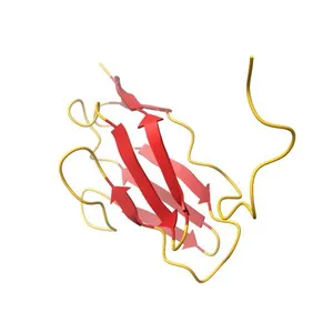
Crystal structure of the C1 domain of cardiac isoform of myosin binding protein-C at 1.3A
Model details
- Platform
NIH 3D
- Price
- Free
- License
- CC-BY
- Print, modify and use commercially — credit the author.
- Formats
- GLBWRLX3DSTL
- 79 Downloads
- 38 Views
Description
PDB Classification: CONTRACTILE PROTEIN
Similar models
Free 
NMR structure of the c3 domain of human cardiac myosin binding protein-c
Free 
NMR structure of the c3 domain of human cardiac myosin binding protein-c with a hypertrophic cardiomyopathy-related mutation R502W.
Free 
Crystal structure of the titin C-terminus in complex with obscurin-like 1
Free 
Solution NMR structure of the C-terminal EF-hand domain of human cardiac sodium channel NaV1.5
Free 
Structure of the immunoglobulin tandem repeat A168-A169 of titin
Free 
High resolution structure of the human alpha-actinin isoform 3
Free 
Crystal structure of the head-to-tail junction of tropomyosin
Free 
Crystal structure of the p53 core domain bound to a full consensus site as a self-assembled tetramer
Free 
Structure of the N-terminal domain of human cardiac troponin C bound to calcium ion and to the inhibitor W7
Free 
Crystal structure of the HIV-1 Rev dimer
Free 
Cardiac human myosin S1dC, beta isoform complexed with Mn-AMPPNP
Names are used only to indicate the source of a result. Polyzon is not affiliated with the listed sites.