Free
3D model · NIH 3D
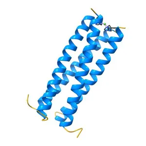
Solution NMR structure of the C-terminal EF-hand domain of human cardiac sodium channel NaV1.5
Model details
- Platform
NIH 3D
- Price
- Free
- License
- CC-BY
- Print, modify and use commercially — credit the author.
- Formats
- X3DWRLGLBSTL
- 78 Downloads
- 36 Views
Description
PDB Classification: METAL BINDING PROTEIN
Similar models
Free
Structure of the N-terminal domain of human cardiac troponin C bound to calcium ion and to the inhibitor W7
Free
Human p53 core domain with hot spot mutation R249S and second-site suppressor mutations H168R and T123A
Free
3D NMR structure of domain cC0 of cardiac myosin binding protein C (MyBPC)
Free
Solution fold of HIV-1 Virus protein U cytoplasmic domain in the presence of DPC micelles
Free
Crystal structure of the C1 domain of cardiac isoform of myosin binding protein-C at 1.3A
Free
Solution NMR Structure of apo-calmodulin in complex with the IQ motif of Human Cardiac Sodium Channel NaV1.5
Free
Solution structure of full-length apo mammalian calmodulin bound to the IQ motif of the human voltage-gated sodium channel NaV1.2
Free
Structural Basis for Substrate Fatty-Acyl Chain Specificity: Crystal Structure of Human Very-Long-Chain Acyl-CoA Dehydrogenase
Free
The Solution Structure of Human Cardiac Troponin C in complex with the Green Tea Polyphenol; (-)-epigallocatechin-3-gallate
Free
Crystal Structure of Human Nav1.4 CTerminal Domain in Complex with apo Calmodulin
Free
NMR structure of calcium-loaded STIM1 EF-SAM
Names are used only to indicate the source of a result. Polyzon is not affiliated with the listed sites.