Free 
3D model · NIH 3D
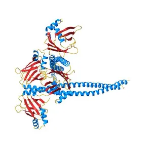
Crystal Structure of Amino Acids 1733-1797 of Human Beta Cardiac Myosin Fused to Gp7
Model details
- Platform
NIH 3D
- Price
- Free
- License
- CC-BY
- Print, modify and use commercially — credit the author.
- Formats
- WRLGLBX3DSTL
- 80 Downloads
- 63 Views
Description
PDB Classification: Actin/DNA Binding Protein
Similar models
Free 
Crystal Structure of Ca2+/CaM:Kv7.4 (KCNQ4) B helix complex
Free 
Crystal structure of the C1 domain of cardiac isoform of myosin binding protein-C at 1.3A
Free 
Crystal structure of the DNA-bound dHax3, a TAL effector, at 1.85 angstrom
Free 
Crystal Structure of the coiled-coil surrounding Skip 3 of MYH7
Free 
Crystal Structure of amino acids 1590-1657 of MYH7
Free 
Crystal structure of mouse myosin-5a in complex with calcium-bound calmodulin
Free 
Crystal structure of IQSEC1-IQ motif, Sec7PH tandem in complex with calmodulin
Free 
Crystal Structure of Amino Acids 1631-1692 of MYH7
Free 
Crystal structure of the TAL effector dHax3 with NI RVD at 2.8 angstrom resolution
Free 
Crystal Structure of Amino Acids 1562-1622 of MYH7
Free 
Structural basis of NR4A1 bound to the human pituitary proopiomelanocortin gene promoter
Names are used only to indicate the source of a result. Polyzon is not affiliated with the listed sites.