Free 
3D model · NIH 3D
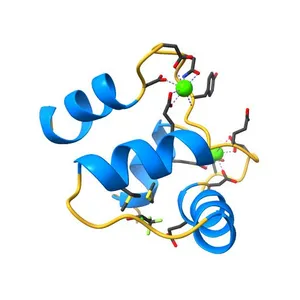
Solution Structure of Ca2+/Calmodulin bound to the C-terminal Domain of Petunia Glutamate Decarboxylase
Model details
- Platform
NIH 3D
- Price
- Free
- License
- CC-BY
- Print, modify and use commercially — credit the author.
- Formats
- X3DGLBWRLSTL
- 78 Downloads
- 55 Views
Description
PDB Classification: BINDING PROTEIN/HYDROLASE
Similar models
Free 
Crystal Structure of Calmodulin Binding Domain of Orai1 in Complex with Ca2+/Calmodulin Displays a Unique Binding Mode
Free 
NMR solution structure of a Ca2+-Calmodulin with a binding motif (NSCaTE) peptide from the N-terminal cytoplasmic domain of the L-type Voltage-Cated Calcium Channel alpha1C subunit
Free 
NMR solution structure of the Ca2+-Calmodulin C-terminal domain in a complex with a peptide (NSCaTE) from the L-type Voltage-Gated Calcium Channel alpha1C subunit
Free 
Solution structure of Magnesium-bound form of calmodulin C-domain E104D/E140D mutant
Free 
Solution NMR-derived structure of calmodulin bound with ER alpha peptides
Free 
Crystal Structure of Calcium-loaded Calmodulin in complex with STRA6 CaMBP2-site peptide.
Free 
Halothane binds to druggable sites in calcium-calmodulin: Solution Structure of halothane-CaM N-terminal domain
Free 
Solution NMR Structure of apo-calmodulin in complex with the IQ motif of Human Cardiac Sodium Channel NaV1.5
Free 
Solution structure of Ca2+/calmodulin complexed with a peptide representing the calmodulin-binding domain of L-selectin
Free 
Structural basis of the Munc13-1/Ca2+-Calmodulin interaction: A novel 1-26 calmodulin binding motif with a bipartite binding mode
Free 
Crystal structure of the adenylyl cyclase domain of anthrax edema factor (EF) in complex with calmodulin and 3' deoxy-ATP
Names are used only to indicate the source of a result. Polyzon is not affiliated with the listed sites.