Free 
3D model · NIH 3D
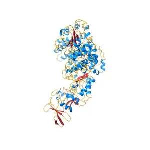
Crystal structure of the adenylyl cyclase domain of anthrax edema factor (EF) in complex with calmodulin and 3' deoxy-ATP
Model details
- Platform
NIH 3D
- Price
- Free
- License
- CC-BY
- Print, modify and use commercially — credit the author.
- Formats
- WRLGLBX3DSTL
- 84 Downloads
- 80 Views
Description
PDB Classification: TOXIN,LYASE/METAL BINDING PROTEIN
Similar models
Free 
Crystal Structure of the EF3-CaM complexed with PMEApp
Free 
Crystal Structure analysis of the adenylyl cyclaes catalytic domain of adenylyl cyclase toxin of Bordetella pertussis in presence of c-terminal calmodulin
Free 
Crystal structure of calmodulin in complex with KN93 (1:1 complex)
Free 
Crystal Structure of Calmodulin Binding Domain of Orai1 in Complex with Ca2+/Calmodulin Displays a Unique Binding Mode
Free 
Crystal structure of the methylated N-ADA/DNA complex
Free 
Crystal structure of a small conductance potassium channel splice variant complexed with calcium-calmodulin
Free 
Crystal Structure of Calcium-loaded Calmodulin in complex with STRA6 CaMBP2-site peptide.
Free 
3D Structure of Calmodulin and Calmodulin binding domain of Olfactory cyclic nucleotide-gated ion channel complex
Free 
X-ray crystal structure of Methylorubrum extorquens AM1 lanmodulin (LanM) with neodymium (III) bound at pH 7
Free 
X-ray Structure of a Core U2AF65/U2AF35 Heterodimer
Free 
STRUCTURE OF A FLAVIN-BINDING DOMAIN, LOV2, FROM THE CHIMERIC PHYTOCHROME/PHOTOTROPIN PHOTORECEPTOR PHY3
Names are used only to indicate the source of a result. Polyzon is not affiliated with the listed sites.