Free
3D model · NIH 3D
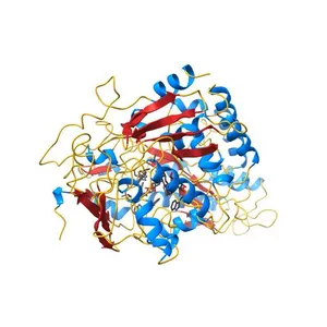
Thermotoga maritima maltotriose binding protein, ligand free form
Model details
- Platform
NIH 3D
- Price
- Free
- License
- CC0
- Public domain: print, modify and sell, no attribution required.
- Formats
- WRLGLBX3DSTL
- 8 Downloads
- 204 Views
Description
PDB Classifiation: SUGAR BINDING PROTEIN
Similar models
Free
Structure and RNA binding of the mouse Pumilio-2 Puf Domain
Free
The periplasmic binding protein component of the arabinose ABC transporter from Shewanella sp. ANA-3 bound to alpha and beta-L-arabinofuranose
Free
Structure of the PDZ domain of human PDLIM2 bound to a C-terminal extension from human beta-tropomyosin
Free
MppA Periplasmic Murein Tripeptide Binding Protein, Unliganded Open Form
Free
Structure of the yeast nuclear exosome component, Rrp6p, reveals an interplay between the active site and the HRDC domain; Protein in complex with Mn
Free
Crystal structure of alcohol dehydrogenase superfamily protein (NP_718042.1) from Shewanella oneidensis at 1.55 A resolution
Free
Crystal structure of cyclic alpha-maltosyl-1,6-maltose binding protein from Arthrobacter globiformis
Free
Crystal structure of the human galectin-4 N-terminal carbohydrate recognition domain in complex with lactose-3'-sulfate
Free
Crystal structure of a dimeric ferredoxin-like protein (bb1511) from bordetella bronchiseptica rb50 at 1.90 A resolution
Free
Sugar binding protein - human galectin-2
Free
Crystal Structure of Monobody CS1/SHP2 C-SH2 Domain Complex
Names are used only to indicate the source of a result. Polyzon is not affiliated with the listed sites.