Free 
3D model · NIH 3D
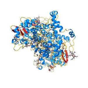
X-ray crystal structure of arachidonic acid bound in the cyclooxygenase channel of G533V murine COX-2
Model details
- Platform
NIH 3D
- Price
- Free
- License
- CC0
- Public domain: print, modify and sell, no attribution required.
- Formats
- WRLGLBSTLX3D
- 8 Downloads
- 149 Views
Description
PDB Classifiation: OXIDOREDUCTASE
Similar models
Free 
The Crystal Structure of Salicylate Bound to Human Cyclooxygenase-2
Free 
Crystal structure of the human beta2 adrenergic receptor in complex with the neutral antagonist alprenolol
Free 
The structure of Ibuprofen bound to cyclooxygenase-2
Free 
Crystal structure of the CXCR4 chemokine receptor in complex with a cyclic peptide antagonist CVX15
Free 
Crystal structure of Phl p 4, a grass pollen allergen with glucose dehydrogenase activity
Free 
Crystal structure of Glucagon-Like Peptide-1 in complex with the extracellular domain of the Glucagon-Like Peptide-1 Receptor
Free 
Crystal structure of tetrameric fibrinogen-like recognition domain of FIBCD1 with N,N'-diacetyl chitobiose ligand bound
Free 
Crystal structure of Tyrosinase from Bacillus megaterium
Free 
The crystal structure of unmodified tRNAPhe from Escherichia coli
Free 
Crystal structure of a human cytochrome P450 2B6 (Y226H/K262R) in complex with the inhibitor 4-(4-Nitrobenzyl)pyridine.
Free 
Human Cytochrome P450 2A6 in complex with nicotine
Names are used only to indicate the source of a result. Polyzon is not affiliated with the listed sites.