Free 
3D model · NIH 3D
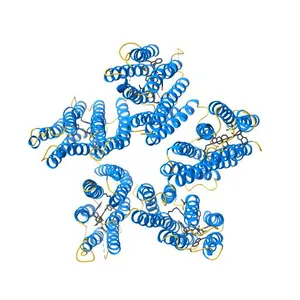
Crystal structure of blue light-absorbing proteorhodopsin from Med12 at 2.3 Angstrom
Model details
- Platform
NIH 3D
- Price
- Free
- License
- CC-BY
- Print, modify and use commercially — credit the author.
- Formats
- X3DGLBWRLSTL
- 10 Downloads
- 100 Views
Description
PDB Classifiation: PROTON TRANSPORT
Similar models
Free 
Crystal structure of a blue-light absorbing proteorhodopsin mutant D97N from HOT75
Free 
Crystal structure of the urea channel from the human gastric pathogen Helicobacter pylori
Free 
Crystal structure of Mn2+ bound calprotectin
Free 
Crystal structure of the quaternary complex of histone H3-H4 heterodimer with chaperone ASF1 and the replicative helicase subunit MCM2
Free 
Crystal structure of KPT185 in complex with CRM1-Ran-RanBP1
Free 
Crystal structure of human ATAD2 bromodomain in complex with 8-2-(dimethylamino)ethylamino-3-methyl-1,2-dihydroquinolin-2-one
Free 
Crystal structure of the class A extended-spectrum beta-lactamase PER- 2
Free 
Crystal structure of M. tuberculosis in complex with BTO - covalent adduct
Free 
Crystal structure of FliG-FliM complex from H. pylori
Free 
Crystal structure of M. tuberculosis TreS
Free 
Crystal Structure of the Cell-Free Synthesized Membrane Protein, Acetabularia Rhodopsin I, at 1.80 angstrom
Names are used only to indicate the source of a result. Polyzon is not affiliated with the listed sites.