Бесплатно Crystal structure of the dimerization domain of Lsr2 from Mycobacterium tuberculosis in the P 1 21 1 space group NIH 3D danielaschipper
Бесплатно human MutSalpha (MSH2/MSH6) bound to a G T mispair, with ADP bound to MSH2 only NIH 3D kitz3d
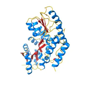
Бесплатно The periplasmic binding protein component of the arabinose ABC transporter from Shewanella sp. ANA-3 bound to alpha and beta-L-arabinofuranose NIH 3D reymeherman
Бесплатно Crystal structure of C-terminal xRRM domain of human Larp7 bound to 7SK stem-loop 4 RNA NIH 3D Andre.cari
Бесплатно Crystal structure of human ribonucleotide reductase 1 bound to the effector TTP NIH 3D gmperez
Бесплатно Crystal structure of cyclic alpha-maltosyl-1,6-maltose binding protein from Arthrobacter globiformis NIH 3D Meghan McCarthy