Бесплатно
3D-модель · NIH 3D
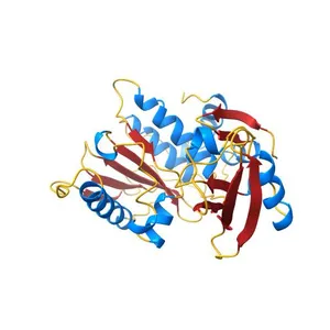
Crystal structure of CYLD USP domain (C596S) in complex with Met1-linked diubiquitin
Сведения о модели
- Площадка
NIH 3D
- Цена
- Бесплатно
- Лицензия
- CC0
- Общественное достояние: можно печатать, изменять и продавать без указания автора.
- Форматы
- X3DGLBWRLSTL
- 8 Загрузки
- 125 Просмотры
Описание
PDB Classifiation: HYDROLASE/PROTEIN BINDING
Похожие модели
Бесплатно
The crystal structure of human Atg4B(C74S)- LC3(1-124) complex
Бесплатно
Structural insights into the interaction of p97 N-terminal domain and SHP motif in Derlin-1 rhomboid pseudoprotease
Бесплатно
Crystal structure of thrombin mutant S195T (free form)
Бесплатно
Crystal Structure of Calcineurin in Complex with the Calcineurin-Inhibiting Domain of the African Swine Fever Virus Protein A238L
Бесплатно
Crystal Structure of Multi-drug Resistant HIV-1 Protease PR-S17 in Complex with Darunavir
Бесплатно
Crystal Structure of sv40 large T antigen origin binding domain with DNA
Бесплатно
Crystal structure of the USP7/HAUSP catalytic domain
Бесплатно
Crystal structure of the C276S mutant of MAP2K7
Бесплатно
Crystal structure of the SARS-CoV-2 (COVID-19) main protease in complex with inhibitor UAW246
Бесплатно
Structure of Florigen Activation Complex Consisting of Rice Florigen Hd3a, 14-3-3 Protein GF14 and Rice FD Homolog OsFD1
Бесплатно
Crystal structures of aldehyde deformylating oxygenase from Oscillatoria sp. KNUA011
Названия используются только для указания источника результата. Polyzon не аффилирован с перечисленными площадками.
Комментарии