Бесплатно 
3D-модель · NIH 3D
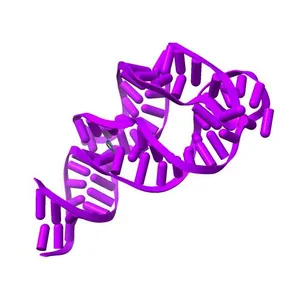
Structure of the adenine riboswitch aptamer domain in an intermediate-bound state
Сведения о модели
- Площадка
NIH 3D
- Цена
- Бесплатно
- Лицензия
- CC-BY
- Можно печатать, изменять и использовать в коммерции — с указанием автора.
- Форматы
- X3DWRLGLBSTL
- 8 Загрузки
- 194 Просмотры
Описание
PDB Classifiation: RNA
Похожие модели
Бесплатно 
Two apo structures of the adenine riboswitch aptamer domain determined using an X-ray free electron laser
Бесплатно 
Crystal Structure of the Domain2 of NAD+ Riboswitch with nicotinamide adenine dinucleotide (NAD+)
Бесплатно 
ATP-bound structure of human platelet phosphofructokinase in an R-state, crystal form II
Бесплатно 
The PROTAC MZ1 in complex with the second bromodomain of Brd4 and pVHL:ElonginC:ElonginB
Бесплатно 
Crystal structure of rhodopsin bound to arrestin by femtosecond X-ray laser
Бесплатно 
Bovine beta-lactoglobulin complex with myristic acid, ambient pressure
Бесплатно 
CRYSTAL STRUCTURE OF THE FMN RIBOSWITCH BOUND TO BRX1151 SPLIT RNA
Бесплатно 
Co-crystal structure of M. tuberculosis ileS T-box in complex with tRNA-3'-OH
Бесплатно 
Crystal structure of the ATP-gated P2X7 ion channel in the closed, apo state
Бесплатно 
pVHL:EloB:EloC in complex with VH298
Бесплатно 
Switching GFP fluorescence using genetically encoded phenyl azide chemistry through two different non-native post-translational modifications routes at the same position.
Названия используются только для указания источника результата. Polyzon не аффилирован с перечисленными площадками.