Бесплатно 
3D-модель · NIH 3D
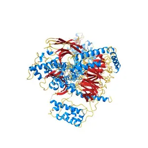
N-Terminal T4 Lysozyme Fusion Facilitates Crystallization of a G Protein Coupled Receptor
Сведения о модели
- Площадка
NIH 3D
- Цена
- Бесплатно
- Лицензия
- CC-BY
- Можно печатать, изменять и использовать в коммерции — с указанием автора.
- Форматы
- WRLGLBX3DSTL
- 9 Загрузки
- 114 Просмотры
Описание
PDB Classifiation: MEMBRANE PROTEIN/HYDROLASE
Похожие модели
Бесплатно 
Crystal Structure of Native Opsin: the G Protein-Coupled Receptor Rhodopsin in its Ligand-free State
Бесплатно 
Crystal structure of the active G-protein-coupled receptor opsin in complex with a C-terminal peptide derived from the Galpha subunit of transducin
Бесплатно 
Crystal structure of the chemokine CXCR4 receptor in complex with a small molecule antagonist IT1t in P1 spacegroup
Бесплатно 
The 2.5 A structure of the CXCR4 chemokine receptor in complex with small molecule antagonist IT1t
Бесплатно 
Structure of active beta-arrestin1 bound to a G protein-coupled receptor phosphopeptide
Бесплатно 
Crystal structure of human calcineurin
Бесплатно 
Structure of the M3 muscarinic acetylcholine receptor bound to the antagonist tiotropium crystallized with disulfide-stabilized T4 lysozyme (dsT4L)
Бесплатно 
Crystal Structure of Calcineurin in Complex with the Calcineurin-Inhibiting Domain of the African Swine Fever Virus Protein A238L
Бесплатно 
Crystal structure of the CXCR4 chemokine receptor in complex with a cyclic peptide antagonist CVX15
Бесплатно 
Mss116p DEAD-box helicase domain 2 bound to a chimaeric RNA-DNA duplex
Бесплатно 
Crystal structure of HIV-1 reverse transcriptase (RT) in complex with Rilpivirine (TMC278, Edurant), a non-nucleoside rt-inhibiting drug
Названия используются только для указания источника результата. Polyzon не аффилирован с перечисленными площадками.