Бесплатно 
3D-модель · NIH 3D
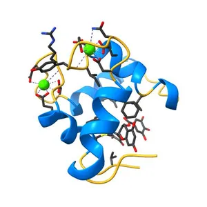
Structure of the N-terminal domain of human cardiac troponin C bound to calcium ion and to the inhibitor W7
Сведения о модели
- Площадка
NIH 3D
- Цена
- Бесплатно
- Лицензия
- CC-BY
- Можно печатать, изменять и использовать в коммерции — с указанием автора.
- Форматы
- GLBX3DSTLWRL
- 78 Загрузки
- 46 Просмотры
Описание
PDB Classification: METAL BINDING PROTEIN
Похожие модели
Бесплатно 
Solution NMR structure of the C-terminal EF-hand domain of human cardiac sodium channel NaV1.5
Бесплатно 
3D NMR structure of domain cC0 of cardiac myosin binding protein C (MyBPC)
Бесплатно 
Crystal structure of the C1 domain of cardiac isoform of myosin binding protein-C at 1.3A
Бесплатно 
NMR structure of the C-domain of troponin C bound to the anchoring region of troponin I
Бесплатно 
NMR structure of the dilated cardiomyopathy mutation G159D in troponin C bound to the anchoring region of troponin I
Бесплатно 
Halothane binds to druggable sites in calcium-calmodulin: Solution Structure of halothane-CaM N-terminal domain
Бесплатно 
Human p53 core domain with hot spot mutation R249S and second-site suppressor mutations H168R and T123A
Бесплатно 
REGULATORY DOMAIN OF HUMAN CARDIAC TROPONIN C IN THE CALCIUM-SATURATED STATE, NMR, 40 STRUCTURES
Бесплатно 
2.0 Angstrom crystal structure of Ca/CaM N53I:RyR2 peptide complex
Бесплатно 
NMR solution structure of a Ca2+-Calmodulin with a binding motif (NSCaTE) peptide from the N-terminal cytoplasmic domain of the L-type Voltage-Cated Calcium Channel alpha1C subunit
Бесплатно 
Halothane binds to druggable sites in calcium-calmodulin: Solution structure of halothane-CaM C-terminal domain
Названия используются только для указания источника результата. Polyzon не аффилирован с перечисленными площадками.