
Grátis 
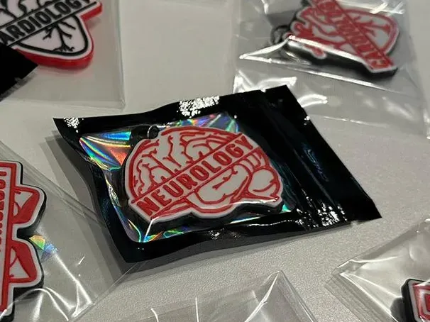
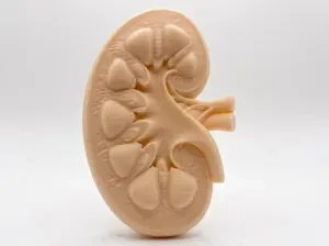
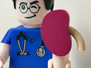
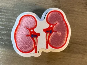
Nephrology Badge - Anatomical Kidney Keychain
MakerWorld
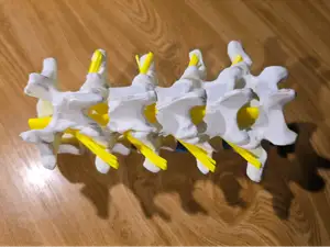
This collection gathers 3D models tied to neurology: brain and ventricular anatomy, neuron and nerve cell figures, cross-sections, and aids used in clinical teaching. Most files are meant for 3D printing at desk or classroom scale, so they work as study objects rather than display pieces alone. It is a practical starting point if you want an STL of a nervous system structure and are not sure which version to pick. Polyzon does not confirm whether models are free, their price, license, official status, affiliation, safety or fitness. Check the current terms at the source.
O texto desta coleção está em inglês; a busca funciona em qualquer idioma.
 Uma coleção do Polyzon · 32 modelos de 6 sites
O Polyzon monta esta coleção a partir dos resultados de busca dos sites conectados. Repetições e modelos sem prévia são removidos, correspondências fracas vão para o final e o restante segue a ordem da busca. Os modelos pertencem aos seus autores — confira os termos na página de cada modelo.
Regras de seleção: equipe editorial do Polyzon · Texto atualizado em 13/10/2026

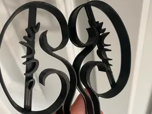


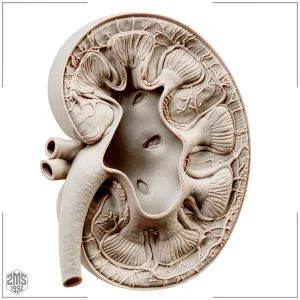









The models fall into a few groups. Whole-brain and ventricular shapes are usually the largest and often come split into halves or paired with a stand, while neuron and nerve cell figures are thinner and more delicate. Clinical aids, such as examination cubes and convergence rules, are flat or geometric and behave differently on the plate than anatomical forms. Cross-section slices and spinal cord pieces sit in between: compact, but with internal detail that decides how they print.
Before printing, open the author's model page and read what is listed there. Check the stated size and scale, how many parts the file contains and whether they are meant to be glued, and whether the author notes that supports are needed. Look at the material and settings the author used, and at photos of real prints, since thin dendrites or hollow ventricles can behave differently in resin and filament. Wall thickness and orientation matter more here than on bulky decorative models.
If the first search feels narrow, try synonyms and related wording: neuroanatomy, cerebral, nerve cell, neuron, spinal cord, brain slice, histology, teaching aid. Related topics such as anatomy, medical education and laboratory accessories often surface models that a single term misses. Combining a structure name with a format or use word usually narrows the list faster than browsing the whole category.
O catálogo completo de temas — de casa e utilidades a CNC, com busca em todos os sites conectados.